
Abstract dark teal wave lines on a solid black background.
Ellipse

At NervGen, the Solution Lies in the Extracellular Matrix

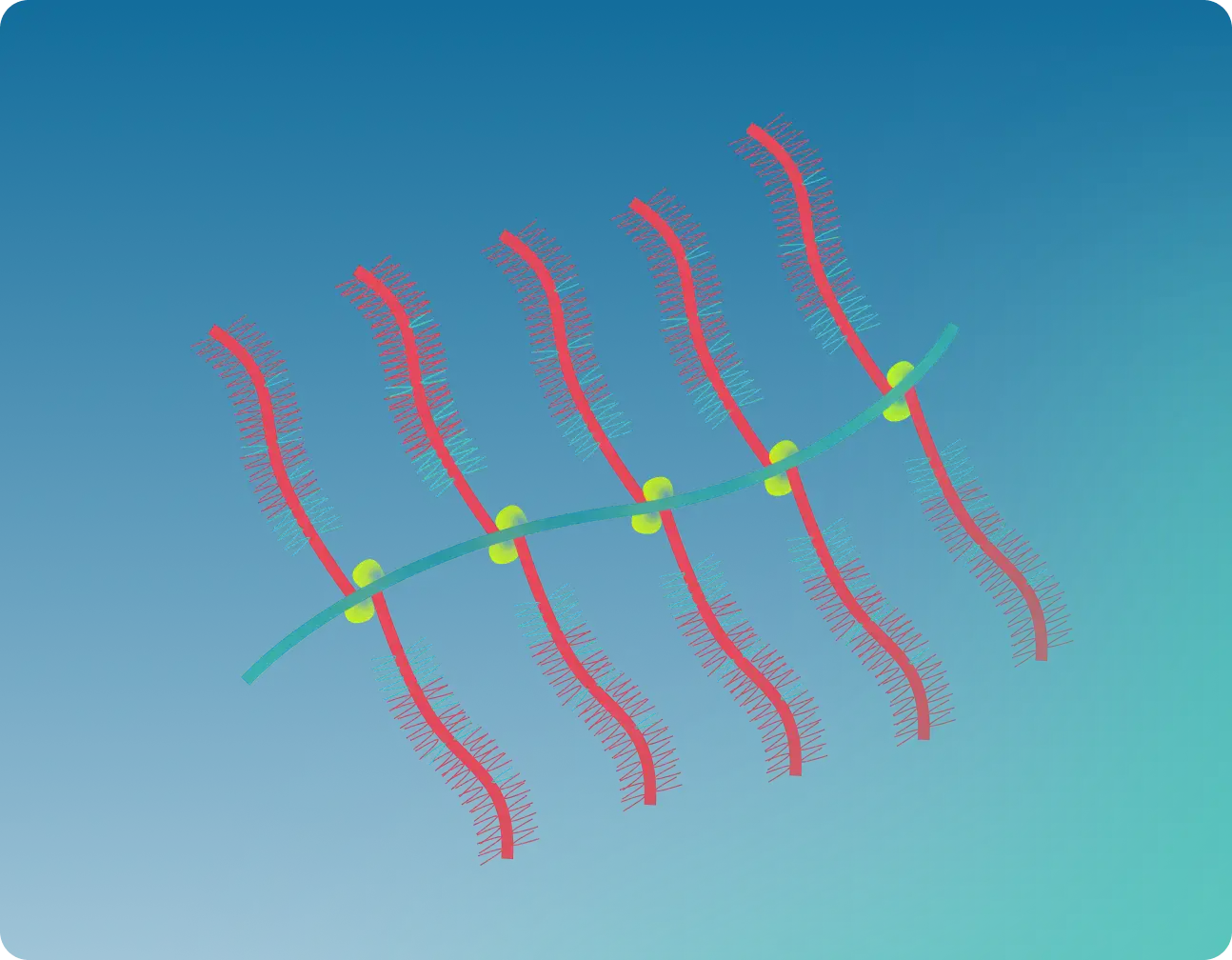
Diagram illustrating an aggrecan proteoglycan structure with labeled hyaluronic acid backbone, core protein, link protein, chondroitin sulfate, and keratan sulfate.
Core Protein (ACAN)
vector arrow.
Link protein
vector arrow.
Chondroitin sulfate
vector arrow.
Keratan sulfate
vector arrow.
Hyaluronic acid backbone
vector arrow.

Chondroitin Sulfate Proteoglycans (CSPGs)

During development of the nervous system, CSPGs guide the formation of neural connections. As the central nervous system matures, they settle into perineuronal nets, specialized structures that stabilize synaptic activity.

After injury, however, CSPG production surges. What once helped build the nervous system now prevents it from repairing itself.

The Glial Scar

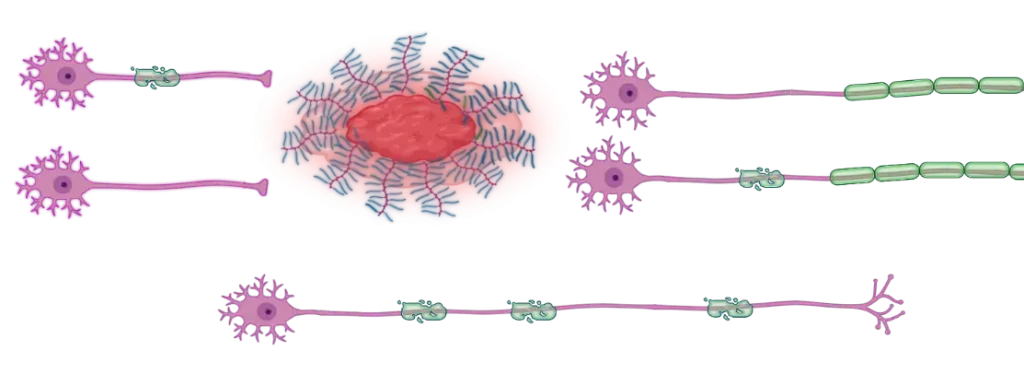
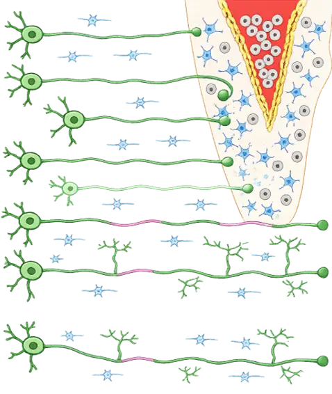
After nervous system damage, the body mounts a defense. Reactive astrocytes form a glial scar, a dense molecular barrier around the damaged tissue.
The protective nature of the glial scar comes at a long-term cost, upregulating the production of CSPGs, an inhibitor of axonal growth.
NVG-291: A New Class of Medicine

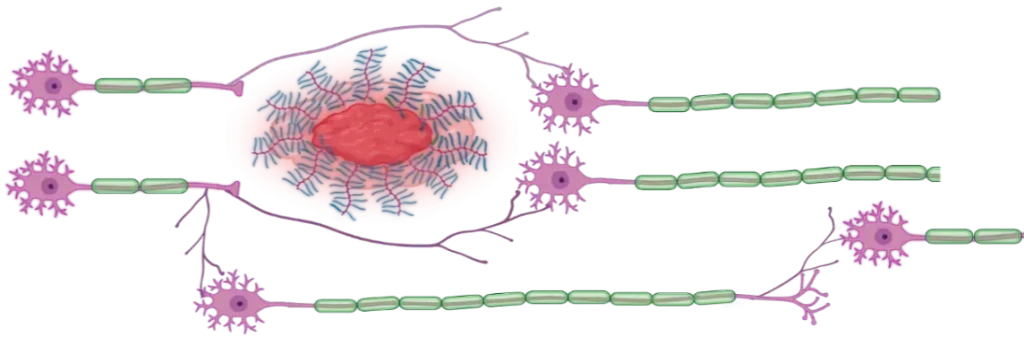
The key to modulating CSPG inhibition lies in the neuronal receptor protein tyrosine phosphatase sigma (PTPσ). NVG-291 is a first-in-class peptide mimetic of the PTPσ wedge domain, designed to unlock the nervous system’s ability to reform neural connections and restore lost function.
Before NVG-291
CSPGs
vector arrow
+ NVG-291
After NVG-291
CSPGs
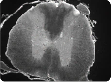
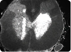
Thoracic & Lumbar 5-HT Staining at 12 Weeks Post-Injury
Thoracic Spinal Cord: Axonal Sprouting
Vehicle
Lesion
Above Injury
Below Injury
NVG-291-R
Lesion
A greyscale cross-section micrograph of a spinal cord, showing the characteristic internal butterfly-shaped gray matter surrounded by white matter tracts.
Vehicle
A greyscale cross-section micrograph of a spinal cord, showing the characteristic internal butterfly-shaped gray matter surrounded by white matter tracts
NVG-291-R
Control
NVG-291-R Treated
blank background.

Beyond Spinal Cord Injury

Abstract blue and teal wave pattern forming an infinity symbol on a white background.

The CSPG-PTPσ pathway is not unique to spinal cord injury. CSPG upregulation is conserved across a wide range of neurotraumatic injuries and neurologic conditions.

Wherever CSPGs accumulate, they block endogenous repair processes. This conserved mechanism allows for NVG-291’s approach to potentially extend well beyond a single condition as a pipeline in a product.
Publications in Scientific Journals

Chondroitinase ABC digestion of the perineuronal net promotes functional collateral sprouting in the cuneate nucleus after cervical spinal cord injury.

.

Journal of Neuroscience. Massey, J. M. et al., 2006; 26, 4406–4414.

Alterations in chondroitin sulfate proteoglycan expression occur both at and far from the site of spinal contusion injury.

.

Experimental Neurology. Andrews, E.M., Richards, R.J., Yin, F.Q., Viapiano, M.S., Jakeman, L.B., 2012; 235, 174–187.

Quantitative changes in perineuronal nets in development and posttraumatic condition.

.

Journal of Molecular Histology. Lipachev, N. et al., 2019; 50, 203–216.

Reactivation of ocular dominance plasticity in the adult visual cortex.

.

Science. Pizzorusso, T. et al., 2002; 298, 1248–1251.

Chondroitinase ABC promotes selective reactivation of somatosensory cortex in squirrel monkeys after a cervical dorsal column lesion.

.

Proceedings of the National Academy of Sciences. Bowes C., Massey J.M., Burish M., Cerkevich C.M., Kaas J.H., 2012; 109 (7) 2595-2600.

Targeting protein tyrosine phosphatase σ after myocardial infarction restores cardiac sympathetic innervation and prevents arrhythmias.

.

Nature Communications. Gardner R.T., Wang L., Lang B.T., et al., 2015; 6(1):6235.

Disrupting protein tyrosine phosphatase σ does not prevent sympathetic axonal dieback following myocardial infarction.

.

Experimental Neurology. Johnsen D., Olivas A., Lang B., Silver J., Habecker B., 2016; 276:1–4.

Correlation between the High-Frequency Content of the QRS on Murine Surface Electrocardiogram and the Sympathetic Nerves Density in Left Ventricle after Myocardial Infarction: Experimental Study.

.

Journal of Electrocardiology. Sedaghat G., Gardner R.T., Kabir M.M., Ghafoori E., Habecker B.A., Tereshchenko L.G., 2017; 50(3):323-331.

ISP and PAP4 peptides promote motor functional recovery after peripheral nerve injury.

.

Neural regeneration research. Lv, Shi-Qin, and Wutian Wu. Vol. 16,8 (2021): 1598-1605. doi:10.4103/1673-5374.294565.

Modulation of proteoglycan receptor regulates RhoA/CRMP2 pathways and promotes axonal myelination.

.

Neurosci Lett. Yao M, Fang J, Tao W, Feng G, Wei M, Gao Y, Xin W, Li Y, Du S. 2021 Aug 24;760:136079. doi: 10.1016/j.neulet.2021.136079. Epub 2021 Jun 21. PMID: 34166723.

Inhibition of the Proteoglycan Receptor PTPσ Promotes Functional Recovery on a Rodent Model of Preterm Hypoxic-Ischemic Brain Injury.

.

Experimental Neurology, October 6, 2023, 114564.

Alzheimer’s disease pathogenesis is dependent on neuronal receptor PTPσ.

.

bioRxiv. Gu Y., Shu Y., Corona A., et al., 2016; 079806.

Perineuronal net digestion with chondroitinase restores memory in mice with tau pathology.

.

Experimental Neurology. Yang S., Cacquevel M., Saksida L.M., et al., 2015; 265:48-58.

Antibody recognizing 4-sulfated chondroitin sulfate proteoglycans restores memory in tauopathy-induced neurodegeneration.

.

Neurobiology of Aging. Yang S., Hilton S., Alves J.N., et al., 2017; 59:197-209.

Reducing hippocampal extracellular matrix reverses early memory deficits in a mouse model of Alzheimer’s disease.

.

Acta Neuropathologica Communications. Végh M.J., Heldring C.M., Kamphuis W., et al., 2014; 11.

Modulation of proteoglycan receptor PTPσ enhances MMP-2 activity to promote recovery from multiple sclerosis.

.

Nature Communications. Luo F., Tran A.P., Xin L., et al., 2018; 9(1):1-16.

Modulating proteoglycan receptor PTPσ using intracellular sigma peptide improves remyelination and functional recovery in mice with demyelinated optic chiasm.

.

Molecular and Cellular Neuroscience. Niknam P., Raoufy M.R., Fathollahi Y., Javan M., 2019; 99:103391.

LAR and PTPσ receptors are negative regulators of oligodendrogenesis and oligodendrocyte integrity in spinal cord injury.

.

Glia. Dyck S., Kataria H., Akbari‐Kelachayeh K., Silver J., Karimi‐Abdolrezaee S., 2018; 67(1):125-145.

Enhanced regeneration and functional recovery after spinal root avulsion by manipulation of the proteoglycan receptor PTPσ.

.

Scientific Reports. Li H., Wong C., Li W., et al., 2015; 5(1):1-14.

Rapid and robust restoration of breathing long after spinal cord injury.

.

Nature Communications. Warren et al., 2018; 9:4843.

Abstract blue and teal wave pattern forming an infinity symbol on a white background.

The Future of Neurorepair Has Begun.
Be Part of What Comes Next.